How the heart pumps blood
Transcript
A fist-sized muscle moves your whole blood supply every minute.
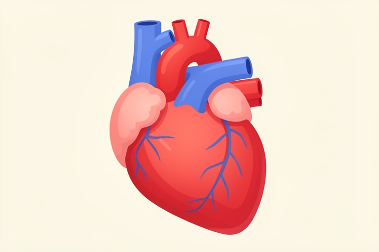
Here is the real heart: four chambers, the atria on top and the ventricles below, each side working as its own pump.
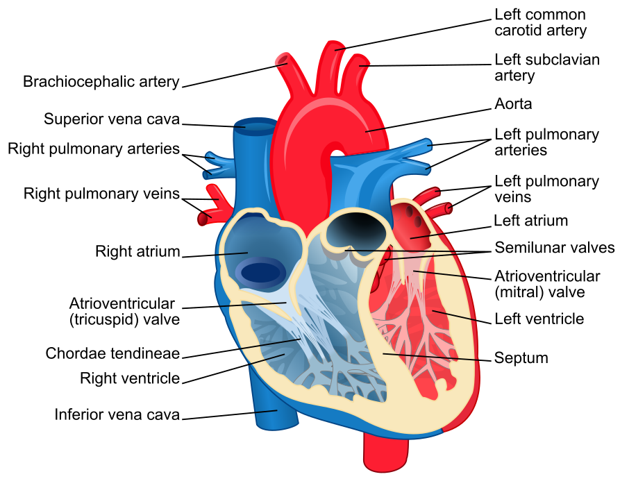
The heart sits in the mediastinum between the lungs, wrapped in a fluid-filled sac called the pericardium.

Its wall has three layers: the outer epicardium, the thick muscular myocardium, and the smooth inner endocardium.
Blood must travel a double circuit: the right heart sends it to the lungs, the left heart sends it to the body.
Each beat has two phases: diastole, when the ventricles relax and fill, and systole, when they contract and eject.


Valves enforce one-way flow: they open when pressure behind them is higher, and snap shut when pressure ahead is higher.
This is why valves need no muscle or nerve: pure pressure difference decides, and it is why backflow is blocked. What triggers the squeeze? The sinoatrial node, the natural pacemaker in the right atrium, fires an electrical impulse.
- Flow goes high to low pressure
The short delay at the AV node lets the atria finish emptying into the ventricles before the ventricles squeeze. Typical resting numbers: about seventy beats a minute and about seventy millilitres per beat, giving cardiac output near five litres a minute.
CO = HR \times SV \approx 70 \times 70\,\text{mL} \approx 5\,\text{L/min}Pressure explains the wall thickness: the left ventricle pushes against the whole body's resistance, so its myocardium is much thicker. A common misconception says blood in the heart feeds the heart muscle, but the heart is fed by its own coronary arteries.

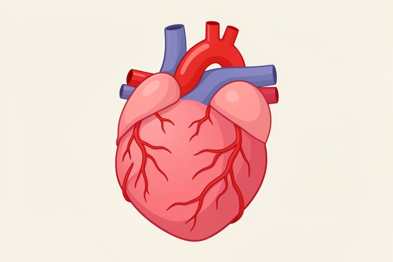
Edge case: if a coronary artery blocks, that muscle starves of oxygen and dies, which is a heart attack. Test your understanding of valves. In short, pressure differences and valves, timed by the pacemaker, keep blood moving one way around two circuits.
- Blocked coronary = heart attack
Quick quiz
Q1. A leaky valve between the left ventricle and aorta lets blood flow backward. What is the main consequence?
Backflow forces rework each beat; the myth is that more blood in the heart means more oxygen for its muscle.